

Views: 0 Author: Maya Medical Clinical Editorial Team Publish Time: 2026-10-27 Origin: Site


Diagnostic imaging represents the clinical backbone of any modern hospital or outpatient diagnostic center. Accurate radiology services enable early disease detection, guide surgical interventions, and reduce unnecessary exploratory procedures. For hospital administrators, chief radiologists, and procurement committees in emerging healthcare markets—particularly across Africa, Southeast Asia, and Central America—investing in radiology equipment requires balancing image resolution, patient throughput, power grid compatibility, radiation safety, and long-term operating costs. This procurement guide outlines the essential technical criteria and clinical considerations for selecting Digital Radiography (DR) X-ray machines, Mobile C-Arm surgical systems, and Color Doppler Ultrasound scanners.
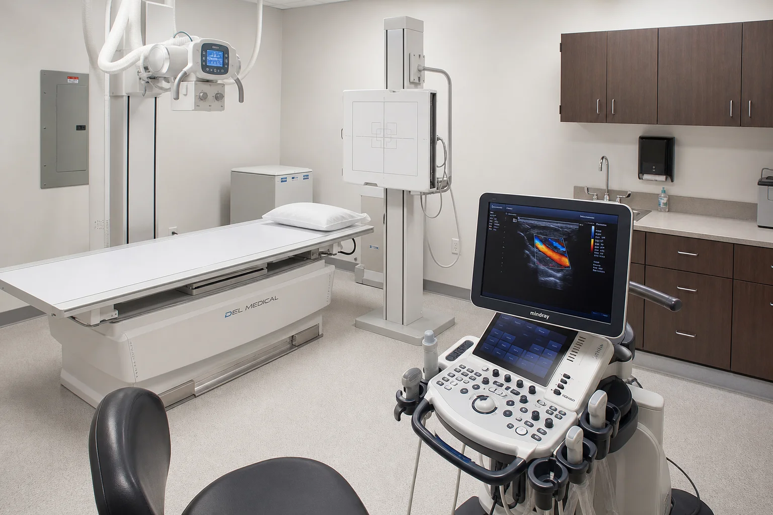
Digital Radiography (DR) has largely supplanted computed radiography (CR) and analog film across modern healthcare facilities due to its superior image acquisition speed, lower radiation dose, and instant digital archiving capabilities. When selecting DR equipment, procurement teams must distinguish between fixed floor-mounted/ceiling-suspended systems and mobile portable units based on clinical case mix and facility architecture.
Fixed DR systems serve as the high-throughput workhorse for outpatient chest, skeletal, abdominal, and trauma imaging. In hospital settings handling 50 to 150 outpatient examinations daily, high-frequency X-ray generators (ranging from 32kW to 50kW or 500mA) provide consistent beam penetration across varying patient body habitus.
U-Arm Multifunctional DR Systems: Highly recommended for facilities with limited floor space (requiring as little as 4m × 3m room dimensions). The motorized U-arm rotates seamlessly between vertical chest views, horizontal table views, and angled decubitus positions without requiring ceiling suspension tracks.
Flat-Panel Detector (FPD) Technology: Facilities should specify Cesium Iodide (CsI) scintillation flat-panel detectors with direct deposition. CsI detectors deliver higher Detective Quantum Efficiency (DQE) and superior spatial resolution (typically 140µm to 100µm pixel pitch) at significantly lower patient X-ray exposure compared to older Gadolinium Oxysulfide (GadOx) alternatives.
For intensive care units (ICUs), emergency trauma bays, and remote field clinics where patients cannot be transported to the radiology suite, mobile digital X-ray units are indispensable. Key procurement requirements include:
Built-in Power Storage: Lithium-ion battery packs capable of sustaining up to 100 to 200 radiographic exposures on a single electrical charge, shielding equipment from local power blackouts.
Wireless Flat-Panel Integration: Wireless 14×17 inch or 17×17 inch FPDs with dedicated anti-drop carbon fiber casing and IPX6 water resistance, communicating via localized Wi-Fi direct to the acquisition console.
Mobile C-Arm fluoroscopy machines provide real-time dynamic X-ray visualization during orthopedic surgery, spine stabilization, trauma reduction, vascular interventions, and urological procedures. Sourcing a C-Arm requires evaluating both mechanical maneuverability and radiation safety management.
Flat-Panel Detector vs. Image Intensifier (II): While older Image Intensifier C-Arms offer lower initial purchase costs, modern Flat-Panel Detector (FPD) C-Arms provide distortion-free rectangular imaging fields, a broader dynamic range (avoiding edge geometric vignette), and up to 40% radiation dose reduction for surgeons and theater nurses.
Generator Power and Anode Thermal Capacity: High-frequency pulsed fluoroscopy generators (minimum 5kW to 15kW) with high-heat-capacity rotating anode X-ray tubes prevent overheating during extended orthopedic and interventional cases.
Orbital Rotation and Free Space: An orbital rotation arc of 135° or greater and a generous source-to-image distance (SID ≥ 100cm) ensures seamless positioning around surgical tables without breaching the sterile surgical drape field.
Ultrasound remains the most cost-effective, radiation-free, and clinically versatile diagnostic tool available to healthcare providers. Modern procurement strategies typically combine high-performance console trolley units for specialized departments with ultra-portable scanners for bedside point-of-care ultrasound (POCUS).
For dedicated obstetrics/gynecology, cardiology, and internal medicine imaging, 4D Color Doppler console systems represent the gold standard. Essential features include:
Transducer Transducer Array Compatibility: Active connectivity for 4 probe ports simultaneously, accommodating high-density convex probes (abdominal/OB), linear probes (vascular/small parts/musculoskeletal), phased array cardiac probes (echocardiography with continuous wave Doppler), and micro-convex transvaginal probes.
Advanced Imaging Algorithms: Speckle reduction imaging (SRI), spatial compound imaging, tissue harmonic imaging (THI), and automated real-time elastography.
For emergency triage, ambulance deployment, rural outreach, and bedside ICU rounds, lightweight laptop-style or wireless handheld ultrasound probes connecting to Android/iOS tablets deliver remarkable clinical diagnostic capability at accessible investment thresholds.
The table below provides a practical procurement checklist for outfitting a comprehensive hospital diagnostic imaging department:
Imaging Modality | Recommended Specification | Target Clinical Application | Critical Facility Requirement |
|---|---|---|---|
Floor-Mounted Digital X-Ray | 50kW / 500mA Generator, CsI Flat Panel (17×17 inch), Motorized Stand | Outpatient chest, skeletal, spine, abdominal radiography | Lead-shielded room (minimum 2.0mm Pb equivalent), 380V/220V stable power |
Mobile Surgical C-Arm | 5kW–10kW Pulsed Generator, Dynamic Flat Panel (21×21cm or 30×30cm) | Orthopedic fixation, trauma surgery, spine procedures | Dedicated surgical OR space, mobile lead aprons & thyroid shields |
Trolley Color Doppler Ultrasound | 15–21 Inch LED Monitor, 4 Active Probe Ports, CW/PW Doppler, 4D OB/GYN | Abdominal, cardiology, obstetrics, vascular, thyroid diagnosis | Temperature-controlled room (20–25°C), acoustic gel supply |
Portable Laptop Ultrasound | Dual-Probe Connectors, Built-in Battery (2–3h runtime), B/M/Color/Power Doppler | Emergency triage, bedside ward rounds, field clinic outreach | Compact storage case, standard AC charging adapter |
Procuring complex diagnostic imaging equipment from overseas requires an experienced manufacturing and biomedical engineering partner. Maya Medical delivers verified radiology systems engineered specifically to withstand challenging clinical and electrical operating environments:
Rigorous Factory Pre-Shipment Calibration: Every fixed digital X-ray generator, mobile C-Arm fluoroscopy unit, and color Doppler ultrasound array undergoes continuous 48-hour burn-in stress testing at Maya Medical's Guangzhou testing facility prior to export dispatch.
Heavy-Duty Voltage & Power Fluctuation Protection: Sensitive digital flat-panel detectors and high-voltage X-ray generators are bundled with industrial-grade servo automatic voltage regulators (AVR) and true online medical UPS units, shielding critical circuits from severe power grid spikes and brownouts.
Factory-Direct Warranty & Spare Parts Lifecycle: Guaranteed 5-to-8-year availability on original X-ray tubes, high-tension cables, digital scintillation flat panels, and ultrasound transducer probe crystals.
Comprehensive Installation & Clinical Training: Real-time bilingual video engineering support via WhatsApp (+86-189-2504-4116), accompanied by full architectural lead-shielding blueprints, wiring schematics, and interactive clinical operation webinars for local hospital radiology staff.
Radiology examination rooms must be lined with lead sheeting or barium sulfate plaster offering a radiation attenuation equivalent of at least 2.0mm to 3.0mm lead (Pb) on all primary beam walls and doors. Control rooms require lead-glass observation windows (minimum 2.0mm Pb equivalent) to ensure complete operator radiation safety.
Cesium Iodide (CsI) crystal needle structures channel X-ray photons directly to the photodiode array with minimal lateral light scattering. This results in significantly higher Detective Quantum Efficiency (DQE) and superior contrast resolution at 30% to 50% lower radiation doses compared to traditional Gadolinium Oxysulfide (GadOx) scintillators.
Maya Medical pre-calibrates all software parameters, detector gain offsets, and tube curves prior to packaging in shock-resistant wooden export crates. Upon delivery, our biomedical engineers connect via live video session to assist local hospital technicians with final positioning, voltage verification, and test phantom exposure verification.


